Mouse antisize:Human CD4 Monoclonal Antibody, 0.1 mg
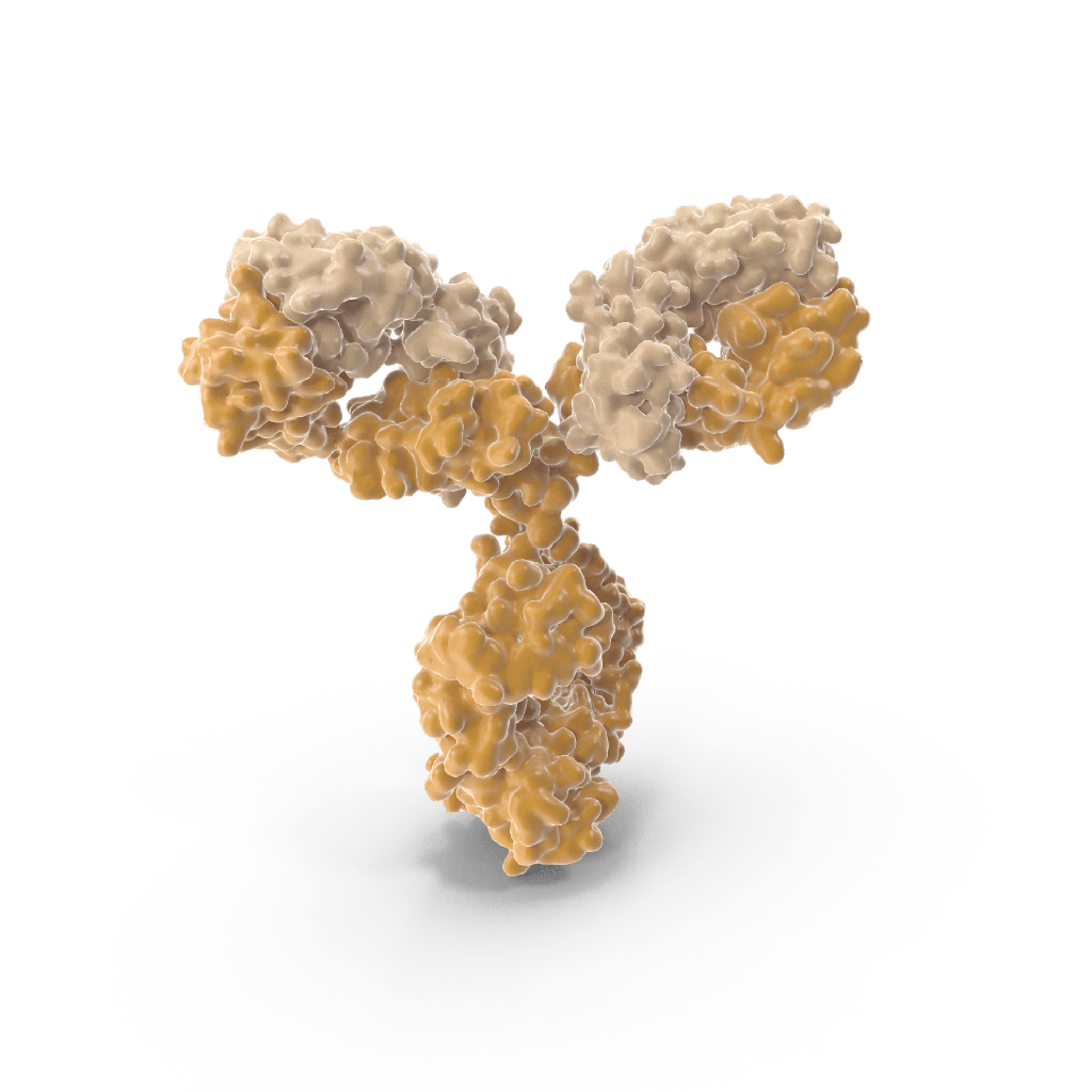
Mouse Anti-Human CD4 Antibody
Introduction
Mouse anti-human CD4 antibody is a monoclonal antibody generated in mice that specifically binds to human CD4 molecules. CD4 is a 55 kDa type I transmembrane glycoprotein expressed on the surface of T-helper lymphocytes, monocytes, macrophages, and dendritic cells. It is encoded by the CD4 gene located on human chromosome 12p13.
Target Protein: Human CD4
CD4 functions as a co-receptor with the T cell receptor (TCR) during antigen recognition by MHC class II molecules. It enhances TCR signaling and participates in the initiation of T-cell activation. CD4 has four extracellular immunoglobulin-like domains (D1–D4), a transmembrane region, and a cytoplasmic tail that interacts with the tyrosine kinase Lck, essential for downstream signal transduction.
Antibody Type
Mouse anti-human CD4 antibodies are typically IgG1 or IgG2a isotype monoclonal antibodies. These antibodies are derived using hybridoma technology by immunizing BALB/c or C57BL/6 mice with recombinant human CD4 protein or CD4+ human cells. Hybridomas producing high-affinity clones are selected for downstream production.
Clone Examples
Common clones include:
- Clone RPA-T4: Frequently used for flow cytometry, binds to domain D1.
- Clone OKT4: Used for immunohistochemistry and flow cytometry.
- Clone SK3: Strong binding affinity, suitable for multicolor cytometry panels.
Each clone differs in epitope specificity, affinity, and functional blocking ability.
Applications
Mouse anti-human CD4 antibodies are widely used in:
- Flow Cytometry (FACS): CD4+ cell enumeration in PBMCs or tissue digests.
- Immunohistochemistry (IHC): Detection of CD4+ T cells in formalin-fixed paraffin-embedded (FFPE) or frozen tissue sections.
- Immunofluorescence (IF): Co-localization of CD4+ T cells in microscopy studies.
- Western Blot (WB): Detecting denatured CD4 in lysates (though not all clones are suitable).
- Cell Sorting (MACS/FACS): Isolation of pure CD4+ T cell populations.
- Functional Blocking: Some clones can block CD4–MHC II interaction for mechanistic studies.
Conjugation Formats
Mouse anti-human CD4 antibodies are available in multiple formats:
- Unconjugated
- Fluorophore-conjugated (e.g., FITC, PE, APC, PerCP-Cy5.5)
- Biotinylated
- HRP-conjugated for ELISA and IHC
- Alexa Fluor® dyes for high-resolution fluorescence microscopy
Host Reactivity
- Species cross-reactivity: Minimal cross-reactivity with non-human primates. No known reactivity with mouse or rat CD4.
- Species specificity: Human-specific, must verify with blocking or isotype controls when working with mixed cell populations.
Storage & Stability
- Recommended storage at 4°C for short-term use.
- Long-term storage at -20°C or below, preferably in aliquots to avoid freeze-thaw cycles.
- Azide and carrier protein content should be checked for compatibility with downstream applications (e.g., functional assays or in vivo use).
Safety and Usage Notes
- Sodium azide in formulations may be toxic for in vivo applications—dialysis recommended.
- When used in blocking or neutralization, titer optimization is required.
- Antibody should be titrated for optimal signal-to-noise in each application.
Certificate of Analysis (COA) Components
Typical COAs include:
- Clone ID and isotype
- Binding specificity by ELISA or FACS
- Endotoxin level (EU/mg)
- Purity (>95%) via SDS-PAGE
- Sterility and mycoplasma testing
Quality Control
Each lot is tested by:
- Flow cytometry using human PBMCs
- ELISA binding assay against recombinant CD4
- IHC staining on human tonsil sections
References and Protocols
- NIH Flow Cytometry Panels: https://www.ncbi.nlm.nih.gov
- Antibody Database: https://antibodyregistry.org
- CD4 protein structure: UniProt P01730
- CD4–MHC II interaction models: Protein Data Bank (PDB ID: 1WIQ)
